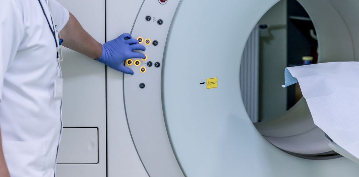
Last time we discussed the limitations of laboratory tests. Today we’ll talk about imaging. You know, x-rays, CT scans, MRIs, Ultrasound, the machines that make pictures of the insides of our bodies. TV shows tell us that if you just order the right scan, it will give the answer we need. The reality is that it’s not that simple. Real life doctors have to weigh many factors to decide on the best approach for each individual patient.
MRIs and CAT scans are not the be all and end all of diagnosis. They can tell us many things about anatomy but they are not perfect. It happens occasionally that surgical findings are different from what a scan showed prior to an operation. Different types of machines also referred to as modalities are used for different purposes and each has its strengths and weaknesses.
A CAT (Computerized Axial Tomography) scan uses x-rays to produce a picture that looks like a slice through the body. It is useful for getting detailed pictures of bones and some soft tissues. It can miss some fractures if they are at the wrong angle and while it can be used to identify some soft tissue masses, it is not well suited for that and can miss them occasionally. A disadvantage is that it uses radiation which, over time and repeated exposure can be detrimental.
MRI (Magnetic Resonance Imaging) is the best modality we have for soft tissue tumors and masses. The machine uses a magnetic field and radio waves to make a picture similar in appearance to a CAT scan. It is commonly used to evaluate the brain and joints. An advantage is that it does not use any dangerous radiation. Some disadvantages are that the machine itself is a bit claustrophobic and it’s probably the most expensive modality and is not easily available everywhere.
Ultrasound is good for fluid filled objects in soft parts of the body close to the skin. It uses technology similar to sonar to make an image with sound waves. This works only in tissues that conduct sound like water and to a lesser degree fat. It works poorly through air filled spaces like the intestines and the lungs. It is usually used to examine pelvic organs and the gallbladder. It doesn’t use radiation and that is why we use it to take pictures of babies. It is not good for looking at structures behind air or bone which don’t transmit sound well. It doesn’t work well for looking deep within the body.
The plain film x-ray is simple, cheap, widely available and has a fair amount of utility. X-rays work well to show bones and things filled with air like lungs or intestines. It doesn’t tell us much about our soft parts and still uses radiation. Mammograms are a special kind of x-ray that is quite difficult to interpret. Radiologists have to look for specific patterns in the films that indicate the possibility of a tumor.
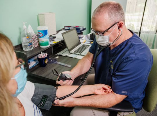
Imaging tests are not perfect. Each has its own idiosyncrasies and no one method is perfect for all situations. They can be quite useful sources of information for figuring out a diagnosis. They are, however, only a part of a larger puzzle that forms as complete a picture as possible of what is happening within a person. The remainder is filled in by the history and input from the patient, physical exam, laboratory testing and the experience and judgement of the physician.


Thanks.